Keynote Lectures
Additional information will be added as it become available, please check back often!
Keynote Lectures are approximately 30 minutes long and cover a wide variety of topics. The 2014 Keynote lecturers will be:
Monday, June 9, 2014
9:30-10:15: Katrin Amunts, Julich Research Center
.jpg) photo source: © Deutscher Ethikrat; Foto: Reiner Zensen |
Katrin Amunts did postdoctoral work in the C. & O. Vogt Institute for Brain Research at Duesseldorf University, Germany. In 1999, she moved to the Research Centre Juelich and set up a new research unit for Brain Mapping. In 2004, she became professor for Structural-Functional Brain Mapping at RWTH Aachen University, and in 2008 a full professor at the Department of Psychiatry, Psychotherapy and Psychosomatics at the RWTH Aachen University as well as director of the Institut of Neuroscience and Medicine (INM-1) at the Research Centre Juelich. In 2013, she became a full professor for Brain Research at the Heinrich-Heine University Duesseldorf, director of the C. and O. Vogt Institute for Brain Research, Heinrich-Heine University Duesseldorf and director of the Institute of Neuroscience and Medicine (INM-1), Research Centre Juelich. |
|
Towards ultra-high resolution models of the human brain |
|
|
Human brain models at microscopical resolution provide the bridge between the cellular level of organization and that of cognitive systems. Data size and complexity of brain organization make it challenging to create them. Models of cellular and fiber architecture were developed based on advanced ICT, opening new perspectives to decode the human brain. |
|
16:15-17:00: Shihui Han, Peking University
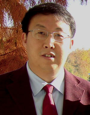 |
Shihui Han is a professor of the Department of Psychology and PKU-IDG/McGovern Institute for Brain Research at Peking University. He is the founding chief editor of Culture and Brain. His research focuses on sociocultural and genetic influences on neural correlates of social cognition such as self-referential processing, empathy for others' pain, and death-related thoughts. |
| Racial In-group Favoritism in Emotion Understanding and Sharing: A Neuroimaging Approach | |
|
Humans understand and share others' emotions and this empathy ability is critical for prosocial behavior. However, we do not empathize everyone equally. Racial in-group favoritism in empathy and altruism has been documented widely. We have been trying to understand the neural correlates of racial in-group bias in empathy using different neuroimaging methods. I'll present the results of our recent neuroimaging studies that outline the neural, cognitive, and genetic mechanisms underlying racial in-group bias in empathy for pain, and different approaches that may reduce the racial in-group bias in empathic neural responses. |
|
Tuesday, June 10, 2014
9:30-10:15: Hanna Damasio, University of Southern California
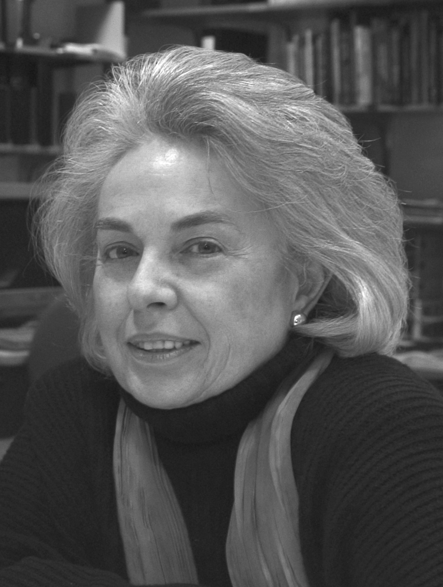 |
Hanna Damasio, a neurologist and cognitive neuroscientist, is University Professor, Dornsife Professor of Neuroscience, and Director of the Dornsife Neuroimaging Center at USC, Los Angeles, and the author of hundreds of scientific articles and an atlas of human brain neuroanatomy based on neuroimaging data. She is a member of the American Academy of Arts and Sciences, the recipient of numerous honorary doctorates, and a highly cited author. |
| Visualizing Human Brain Anatomy | |
|
Early lesion method studies were the forerunners of both cognitive neuroscience and human neuroimaging, a fact often obscured by the hundred years that would pass before modern brain scanning began in the 1970’s. I will review this history in the perspective of human brain anatomy as revealed by current neuroimaging. |
|
16:15-17:00: James Haxby, Director for the Center for Cognitive Neuroscience at Dartmouth and a professor at the Center for Mind/Brain Sciences (CIMeC), University of Trento
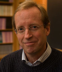 |
Jim Haxby is the Director for the Center for Cognitive Neuroscience at Dartmouth and a professor at the Center for Mind/Brain Sciences (CIMeC), University of Trento. His research in visual neuroscience currently focuses on neural decoding using multivariate pattern analysis (MVPA) and |
| A common high-dimensional linear model of representational spaces in human cortex | |
|
The functional architecture of human cortex can be modeled as high-dimensional representational spaces in which patterns of brain activity are recast as vectors with basis functions that have tuning profiles and patterns of connectivity that are common across brains. Transformation matrices that rotate individual anatomical spaces into the common model space are derived with searchlight-based, whole cortex hyperalignment. Patterns of brain activity in individual brains are modeled as multiplexed topographic basis functions. This model provides a common structure that captures fine-grained distinctions among cortical patterns of response that are not modeled well by current brain atlases. |
|
Wednesday, June 11, 2014
9:30-10:15 Yaniv Assaf, Tel Aviv University
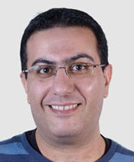 |
Yaniv Assaf completed PhD in chemistry at Tel Aviv University (2001) followed by a post-doctorate at |
| The Role of Neuroimaging in Redefining Neuroplasticity Beyond The Synapse | |
|
Neuro-plasticity is one of the key processes in brain's physiology. In the last decade, structural MRI studies of long-term brain plasticity revealed significant volumetric/regional changes following weeks of training. Yet, none of the known hallmark mechanism of neuroplasticity (i.e. synaptic plasticity) can explain these effects. As a consequence, the micro-structural and cellular correlates of these structural plasticity changes are not well understood. In the lecture, we will explore the origins of structural plasticity, as revealed by MRI, and it's evolution in time (from seconds to months). We will discuss the impact of using MRI in studying neuroplasticity – the ability to localize and explore this basic process of brain physiology, in-vivo and for the whole brain both in rodents and humans. |
|
16:15-17:00 Richard Frackowiak, LREN, Département des Neurosciences Cliniques, CHUV, Université de Lausanne, Lausanne, Switzerland
 |
Richard Frackowiak is Professor and head of the Department of Clinical Neurosciences at the Université de Lausanne (UNIL) and its Centre Hospitalier Universitaire Vaudois (CHUV). He is a co-executive director of the EU’s “Human Brain Project”. He founded the Wellcome Department of Imaging Neuroscience and the FIL in 1994, and has also worked at the ENS in Paris. |
| The Role of Neuroimaging in the Human Brain Project | |
|
Human functional and structural brain imaging with MRI continues to revolutionise tissue characterisation from development, through ageing and as a function of disease. Multi-modal and multi-sequence imaging approaches that measure different aspects of tissue integrity are leading to a rich mesoscopic-level characterisation of brain tissue properties. Novel image classification techniques that capitalise on advanced machine learning techniques and powerful computers are opening the road to individual brain analysis. Data-mining methods, often developed in other data-rich domains of science, especially particle and nuclear physics, are making it possible to identify causes of disease or its expression from patterns derived by exhaustive analysis of combinations of genetic, molecular, clinical, behavioural and other biological data. Imaging is generating data that links molecular and cellular levels of organisation to the systems that subtend, action, sensation, cognition and emotion. These ideas will be illustrated with reference to the human dementias and in the context of the Human Brain Project. |
|
Thursday, June 12
9:30-10:15: John Duncan, Medical Research Council, Cambridge, UK
 |
Educated at the University of Oxford (1970-1976), John Duncan did postdoctoral research with Michael Posner at the University of Oregon, before taking up a position with the Medical Research Council in Cambridge where he has remained ever since. His research extends across neuropsychology, neuroimaging, and single cell electrophysiology, addressing problems of attention, intelligence, and cognitive control. |
|
A core brain system in assembly of cognitive episodes |
|
|
All human cognition is controlled in a series of attentional episodes, breaking complex problems into simpler, more solvable sub-problems. In human fMRI studies, a common or multiple-demand (MD) pattern of frontal and parietal activity is associated with diverse cognitive demands, and with standard tests of fluid intelligence. Based on behavioural, neuropsychological, fMRI and single unit data, I suggest that the core function of MD cortex is to control complex cognition in a structured sequence of attentional episodes. |
|